Introduction
FBA is a relatively common but potentially life-threatening event in children, particularly in those under the age of five [1]. However, in older children, the diagnosis may be delayed or missed due to the absence of a clear history of aspiration and the non-specific nature of clinical and radiological signs [2]. Retained endobronchial foreign bodies can lead to persistent or recurrent pulmonary infections, atelectasis, or even irreversible lung damage if not promptly identified and treated [3].
While flexible or rigid bronchoscopy remains gold standard for both diagnosis and removal, certain cases pose diagnostic and therapeutic challenges, especially when the foreign body is radiolucent or not easily visualized endoscopically [4-6]. Surgical intervention may be necessary when bronchoscopic extraction fails or when complications arise [7].
We report a rare and complex case of a metallic bronchial foreign body in an 11-year-old child, initially missed on bronchoscopy, ultimately requiring surgical bronchotomy after multiple unsuccessful attempts at endoscopic removal. This case underscores the importance of maintaining a high index of suspicion and the value of multidisciplinary collaboration in managing complicated foreign body aspirations in children.
Case presentation
An 11-year-old boy with no significant past medical history presented initially to the pediatric department with symptoms consistent with an infectious pneumonia. Despite receiving appropriate antibiotic therapy, the patient showed no clinical or radiological improvement over several weeks, raising suspicion for an alternative diagnosis. Physical examination revealed decreased breath sounds over the left hemithorax, but no fever or acute respiratory distress was noted at this stage.
A chest Computed Tomography (CT) scan performed on April 19, 2025, identified a metallic foreign body lodged within the bronchus of the left upper lobe. This obstruction was associated with localized pneumonic infiltrates affecting the lingular and anterobasal segments. Notably, no history of choking or witnessed aspiration was reported by the family.
An initial flexible bronchoscopy was conducted in the Ear, Nose, and Throat (ENT) department with the aim of visualizing and potentially extracting the foreign body. However, this procedure failed to identify the object, possibly due to its deep embedment or anatomical complexity. Subsequently, the child was referred to our pulmonology department for further evaluation and management.
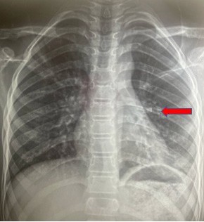
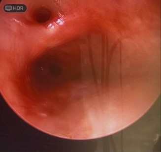
Upon reevaluation, a chest X-ray confirmed the presence of a V-shaped metallic foreign body in the left bronchial tree (Figure 1). A second bronchoscopy, this time performed under general anesthesia in the operating room, revealed an unexpected anatomical variant: A non-catheterizable supernumerary bronchus at the distal end of the left main bronchus. Despite careful exploration, the foreign body remained unseen (Figure 2).
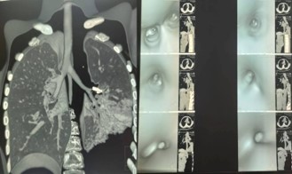
To better characterize the lesion, a chest angio-CT was performed on May 3, 2025. This imaging confirmed the presence of the metallic foreign body embedded within the lingular and culminal bronchi. Additionally, a short segment bronchial stenosis was identified at the origin of the left lower lobe bronchus. The pneumonia showed partial regression, suggesting some improvement under medical therapy.
Virtual bronchoscopy reconstructions were generated from the CT data to aid in procedural planning (Figure 3). After multidisciplinary consultation involving pulmonologists, radiologists, and thoracic surgeons, an attempt was made to remove the foreign body via rigid bronchoscopy. Unfortunately, this intervention was unsuccessful as the foreign body could not be visualized or grasped.
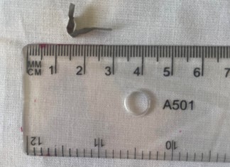
Given the failure of endoscopic extraction, surgical management was undertaken. On July 1, 2025, the patient underwent a bronchotomy of the left main bronchus through a thoracotomy approach. The metallic foreign body was successfully extracted without intraoperative complications (Figure 4).
On postoperative day 2, a follow-up bronchoscopy showed the previously noted supernumerary bronchus to be patent. However, whitish secretions indicative of ongoing inflammation was present in the left main bronchus, along with mucosal erythema and minor bleeding. Clinically, the child remained afebrile and did not report pain. Auscultation revealed a persistent silence over the left lung field.
Subsequent imaging demonstrated worsening, with a “white lung” appearance on chest radiograph and a moderate pleural effusion identified on thoracic ultrasound. On postoperative day 10, a CT-guided drainage using a pig-tail catheter was performed, successfully evacuating 500 mL of clear pleural fluid. The drain was removed after four days following clinical and radiological improvement.
Follow-up chest X-rays confirmed re-expansion of the left lung, with only a small residual apical hydroaeric level. The patient’s clinical course remained favorable, with resolution of symptoms under continued antibiotic therapy and chest physiotherapy.
Discussion
Foreign Body Aspiration (FBA) is a true pediatric emergency, with the potential to rapidly evolve into life-threatening respiratory compromise or insidious chronic pulmonary disease [8,9]. It remains one of the leading causes of accidental death in young children, particularly those under the age of five [9,10]. However, as demonstrated in our case, older children and adolescents are not exempt and may present with more subtle, atypical, or misleading symptoms. These cases represent a diagnostic blind spot, where delayed recognition can lead to significant morbidity.
In younger children, the diagnosis of FBA is often facilitated by a classic triad of symptoms: sudden onset of coughing, choking, and unilateral wheezing [2]. In contrast, older children may not report a clear aspiration event, either because they do not recognize the incident as problematic, or because the symptoms are mild or intermittent [3]. Moreover, as in our case, the absence of respiratory distress or dramatic clinical signs may lead physicians to favor more common diagnoses, such as community-acquired pneumonia. This often results in repeated courses of antibiotics with minimal or no improvement, ultimately delaying the correct diagnosis and appropriate intervention.
A further diagnostic difficulty in this case was the absence of any recalled aspiration event. Even after surgical removal and direct visualization of the metallic foreign body, the child did not recognize or remember having inhaled the object. This strongly suggests that the aspiration was likely chronic or remote, possibly occurring weeks, months or even years prior to presentation. Such scenarios are not uncommon in older children and adolescents, who may not perceive minor choking episodes as significant, or may fail to report them entirely. In cases where the foreign body is small, non-obstructive, or initially well tolerated, symptoms may be mild, intermittent, or entirely absent, contributing to diagnostic delays.
Over time, however, the chronic retention of a foreign object within the airway can induce a local inflammatory response, resulting in mucosal edema, granulation tissue formation, and complications such as recurrent infection, segmental atelectasis, and bronchial stenosis—several of which were present in our patient [11]. This reinforces the need for clinicians to maintain a high index of suspicion for FBA in any child with unexplained or persistent respiratory symptoms, even in the absence of a clear aspiration history.
Radiological imaging is essential in the diagnostic process, but its sensitivity and specificity depend on the nature of the foreign body and the timing of imaging. Chest X-ray remains the first-line investigation, particularly in resource-limited settings. However, up to 50% of aspirated foreign bodies are radiolucent, leading to normal or nonspecific findings such as atelectasis, hyperinflation, or localized infiltrates [11,12]. In our patient, the radio-opaque nature of the metallic foreign body facilitated its detection on plain radiography, but the precise anatomical location, degree of obstruction, and secondary parenchymal changes were far better assessed through high-resolution Computed Tomography (CT).
CT imaging—particularly when combined with angiographic protocols and virtual bronchoscopy—proves invaluable in cases where bronchoscopy is inconclusive. It enables detailed three-dimensional visualization of the tracheobronchial tree and facilitates the detection of associated complications such as bronchial stenosis, abscess formation, or migration of the foreign body [13]. In our case, CT not only confirmed the shape and precise location of the impacted foreign body, but also revealed a short segment stenosis at the origin of the left lower lobe bronchus, along with imaging features consistent with a fully healing granulomatous reaction.
Bronchoscopy, whether flexible or rigid, is considered the diagnostic and therapeutic gold standard. Flexible bronchoscopy offers superior visualization of distal airways and is often used in initial diagnostic workups, especially in older or cooperative children. Rigid bronchoscopy, on the other hand, is preferred in younger children for foreign body extraction due to superior airway control and the availability of a wider range of retrieval instruments [14-16]. However, both techniques have limitations. Visualization can be obscured by granulation tissue, inflammation, purulent secretions, or complex anatomical variants—such as the supernumerary bronchus encountered in our patient. In such cases, repeated unsuccessful bronchoscopies can increase mucosal injury, risk of perforation, and procedural anxiety for both patient and caregivers.
Surgical intervention for FBA is rare, typically reserved for cases where bronchoscopic extraction fails or complications necessitate direct access. It is reported in less than 2% of pediatric FBA cases in large series [17]. Indications for surgery include tightly embedded foreign bodies, significant airway stenosis, bronchial wall perforation, or when the foreign body is in an anatomically inaccessible site. Our patient underwent a left main bronchus bronchotomy after multiple unsuccessful endoscopic attempts. This intervention allowed for the complete removal of the V-shaped metallic object and avoided further risk of chronic infection or airway damage.
Postoperative complications, though uncommon, must be anticipated. In our case, the child developed left lung atelectasis and a moderate pleural effusion, which required intervention. Mucus plugging is a frequent cause of postoperative atelectasis, and bronchoscopy remains the best method for clearance. The pleural effusion, likely reactive and sterile, was successfully drained under CT guidance using a pigtail catheter. The patient responded well to supportive care, antibiotics, and respiratory physiotherapy, with near-complete radiological resolution at follow-up.
This case highlights several key clinical takeaways:
• High clinical suspicion is critical in any child with persistent or recurrent pneumonia, particularly when unresponsive to antibiotics and in the absence of other risk factors.
• Atypical or silent presentation of FBA is common in older children and adolescents and should not be underestimated.
• Advanced imaging —including CT with virtual bronchoscopy—should be considered early in unclear or complicated cases.
• Anatomical variants, such as supernumerary bronchi, though rare, may interfere with diagnostic bronchoscopy and increase the risk of misdiagnosis.
• Surgical extraction, although infrequent, should be pursued without delay in selected cases to avoid prolonged inflammation, bronchial remodelling, or irreversible damage.
Finally, this case underscores the value of a multidisciplinary approach, integrating paediatricians, pulmonologists, radiologists, anaesthesiologists, ENT and thoracic surgeons, and physiotherapists. Timely collaboration allowed for an accurate diagnosis, safe surgical removal, and effective postoperative care, ultimately leading to a full recovery without long-term sequelae.
Conclusion
This case report illustrates the diagnostic and therapeutic challenges posed by Foreign Body Aspiration (FBA) in older children. While classically associated with early childhood, FBA can occur at any age and may present with subtle or atypical symptoms, leading to delayed recognition. In our case, the absence of a clear history of aspiration and the anatomical complexity contributed to multiple failed bronchoscopic attempts.
Advanced imaging modalities such as CT and virtual bronchoscopy were instrumental in identifying the foreign body and guiding further management. When endoscopic approaches failed, surgical intervention via bronchotomy provided a definitive solution, demonstrating the importance of adapting the treatment strategy to the clinical context.
Postoperative follow-up is essential due to the risk of complications such as atelectasis and pleural effusion, which can be effectively managed with bronchoscopy, imaging-guided drainage, and respiratory physiotherapy. A multidisciplinary and individualized approach is key to achieving favourable outcomes in such complex cases.
References
- Louhaichi S, Boubaker N, Hamdi B, Nemsi E, Ouerghi S, Mestiri T, et al. Removal of airway foreign body using flexible bronchoscopy in children. Arch Pediatr. 2024; 31: 264-269.
- Mîndru DE, Păduraru G, Rusu CD, Țarcă E, Azoicăi AN, Roșu ST, et al. Foreign body aspiration in children—retrospective study and management novelties. Medicina (Kaunas). 2023; 59: 1113.
- Lowe DA, Vasquez R, Maniaci V. Foreign body aspiration in children. Clin Pediatr Emerg Med. 2015; 16: 140-148.
- Mansour B, Elias N. Foreign body aspiration in children with focus on the role of flexible bronchoscopy: a 5-year experience. Isr Med Assoc J. 2015; 17: 599-603.
- Costa AS, Palomino ALM, Suzuki I, Garcia RG, Scordamaglio PR, Gregorio MG, et al. Analysis of 108 flexible bronchoscopies for the removal of foreign bodies from the airways. Einstein (Sao Paulo). 2023; 21: eAO0391.
- Shao H, Li S, He J, Wu L, Chen Z. A combination of flexible and rigid bronchoscopy in the successful removal of a residual fish bone from a peripheral bronchus: a case report. Front Pediatr. 2023; 11: 1114043.
- [Authors not listed]. Safety of surgical removal of aspirated foreign bodies after failure of bronchoscopy removal. Curr Respir Med Rev. 2023; 19: 93-96.
- D’Addio E, Palma PL, Di Sessa A, Guarino S, Marzuillo P, Apicella A. Foreign body aspiration in children—diagnostic clues through a clinical case. Pediatr Rep. 2022; 14: 81-85.
- Erdem AO, Etensel B, Yazıcı M, Özkısacık SK, et al. Diagnostic evaluation of foreign body aspiration in children. J Pediatr Res. 2021.
- Passàli D, Lauriello M, Bellussi L, Passali GC, Passali FM, Gregori D. Foreign body inhalation in children: an update. Acta Otorhinolaryngol Ital. 2010; 30: 27-32.
- Shin SM, Kim WS, Cheon JE, Jung AY, Youn BJ, Kim IO, et al. CT in children with suspected residual foreign body in airway after bronchoscopy. AJR Am J Roentgenol. 2009; 192: 1744-1751.
- Rashid M, Saleem H, Ayaz S, Nabi I, Manzoor F. Chest X-ray findings and types of foreign bodies in patients presenting with complaints of tracheobronchial foreign body aspiration: a nine-year multicenter prospective study. J Pak Med Assoc. 2020; 70: 956-961.
- Haliloglu M, Ciftci A, Oto A, Gumus B, Tanyel F, Senocak M, et al. CT virtual bronchoscopy in the evaluation of children with suspected foreign body aspiration. Eur J Radiol. 2003; 48: 188-192.
- Li D, Yang N, Dong J, Wang J, Cui X, Yu H, et al. Comparison of rigid bronchoscopy and flexible bronchoscopy for the management of foreign body aspiration in children. Am J Otolaryngol. 2023; 45: 104092.
- Flexible versus rigid bronchoscopy for tracheobronchial foreign body removal in children: a comparative systematic review and meta-analysis. J Clin Med. 2025; 13: 5652.
- Use of flexible bronchoscopy in foreign body aspiration. Turk Arch Otorhinolaryngol. 2022.
- Elaziz MA, Eltaweel M, Elsayed M, El-Hag-Aly M. Bronchotomy as a safe and feasible alternative to failed bronchoscopic removal of foreign body inhalation in pediatrics: a seven-year review. Open J Thorac Surg. 2021; 1: 47-57.