Introduction
The MUS remains the gold standard for the treatment of Stress Urinary Incontinence (SUI) [1]. While the procedure has low complication rates, significant vascular complications including injury to the obturator, internal iliac and inferior epigastric vessels can be detrimental [2]. Surgeons performing a MUS should be familiar with the regional anatomy and management of such complications [3]. Additionally, studies have shown that complications are less common when these procedures are performed by high-volume surgeons [3]. Retropubic hematomas following MUS placement are usually venous in origin and are estimated to occur in 5-8% of MUS surgeries. These hematomas are often self-limited and active bleeding resolves after 5-10 minutes of direct pressure or application of a hemostatic agent [4]. However, there is a paucity of literature regarding the incidence and management of rectus sheath hematomas following MUS placement. Here, we present a unique case of a delayed, superinfected rectus sheath hematoma following placement of a MUS.
Case
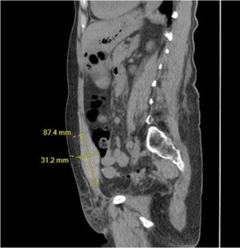
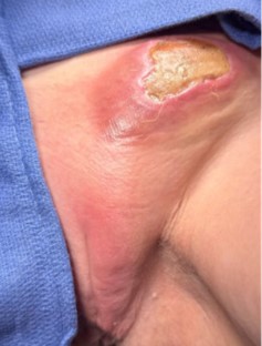
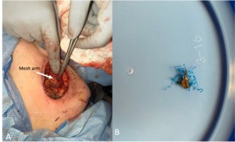
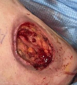
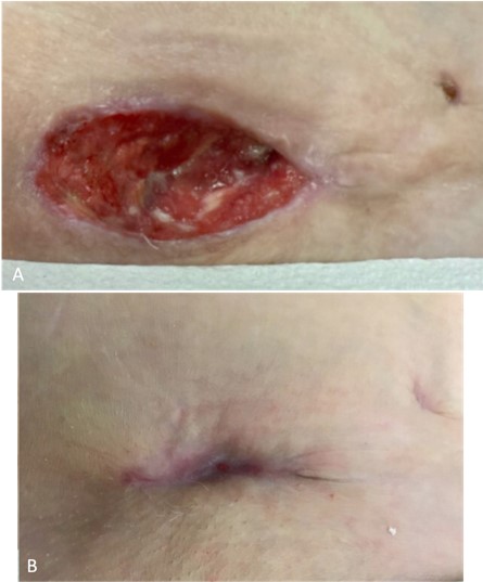
The patient is a 73-year-old woman with a past medical history of hypertension who underwent an uncomplicated, isolated MUS surgery for bothersome SUI. One week following her procedure, she presented to the local emergency room due to concerns for an infection surrounding the left suprapubic MUS trocar site and shortness of breath. Her labs were notable for a creatinine of 5.4 and lactate of 3.2. An echocardiogram revealed ischemic cardiomyopathy of unknown etiology with an ejection fraction of 25%. A Computed Tomography (CT) demonstrated a 4.3×3.0×8.7 cm left-sided rectus sheath hematoma (Figure 1). She was admitted for sepsis secondary to cellulitis of the left MUS trocar site and was initiated on IV piperacillin/tazobactam and linezolid. After one week, her laboratory values and symptoms improved, and she was discharged with a course of oral doxycycline. Her blood and urine cultures obtained during admission were negative. She represented ten days later with increasing pain and purulent drainage from the left suprapubic trocar site. Her laboratory evaluation revealed an acute kidney injury and a urinary tract infection. Repeat CT demonstrated a stable rectus sheath hematoma. An echocardiogram showed an improved ejection fraction of 41%. She was initiated on IV imipenem-cilastatin, and general surgery and Ob/Gyn were consulted. Both surgical teams recommended transfer to a tertiary care center, and she was subsequently transferred to our institution. On arrival, she was nontoxic and afebrile. Her labs were notable for a lactate of 2.4 and a hemoglobin of 8.0. Her white blood cell count was normal. On exam, the left MUS trocar site had a 4.0×3.0 cm, erythematous, non-purulent eschar with significant firmness, along with erythema and induration extending down the left labium majus. Vaginal exam was non-tender, with no evidence of retropubic or vaginal sidewall hematoma. Her vaginal incision was intact and well-healed. Infectious disease and internal medicine were consulted, and she was continued on empiric antibiotics. Given her significant medical comorbidities, surgical exploration with debridement was deferred and the patient underwent ultrasound guided drainage of the hematoma with Interventional Radiology (IR). Minimal sanguineous fluid was aspirated, and outpatient follow up with her local wound clinic was planned. She was discharged on oral amoxicillin/clavulanic acid which was later discontinued as both hematoma aspirate and blood cultures were negative. Two weeks later, she represented to our institution due to continued concerns about the left MUS trocar site. Laboratory evaluation was negative for infection. CT showed resolution of the hematoma, however new subcutaneous fat stranding along the left trocar site, extending down to the left labium majus was noted. Her exam demonstrated a nonhealing left trocar wound with new skin necrosis and continued induration of the left labium majus without fluctuance (Figure 2). General surgery was consulted, and the decision was made to proceed with exam under anesthesia, incision and debridement of the abdominal wound and cystourethroscopy with urogynecology and general surgery. Intraoperatively, the wound was opened, explored, and debrided. The left trocar site was probed and tracked down to the left labium majus. Necrotic tissue was removed and sent to pathology. The distal portion of the left mesh arm was encountered during debridement (Figure 3A), and the small amount of mesh above the level of the fascia was trimmed and sent to pathology (Figure 3B). Following debridement, healthy tissue was exposed (Figure 4). A Penrose drain was left in place while admitted postoperatively, and she continued to improve with empiric antibiotic therapy. Intraoperative wound cultures resulted with Citrobacter freundii, Enterococcus faecalis and Staphylococcus epidermidis. Her final tissue pathology demonstrated gangrenous necrosis, negative for infection. On postoperative day five, the Penrose drain was removed, she was transitioned to oral antibiotics and discharged with home health for twice daily wet to dry dressing changes. In addition to close monitoring with home health, she was followed outpatient by urogynecology and general surgery. Her postoperative course was uncomplicated, and the progression of wound healing is shown in (Figure 5).
Discussion and Conclusion
Rectus sheath hematomas can result from injury to the inferior epigastric vessels or to the rectus muscle itself [5]. While unusual, delayed large vascular injuries following MUS have been reported [6]. Here, we present a delayed rectus sheath hematoma one week after an uncomplicated, isolated MUS placement. To our knowledge, this is the first case report detailing this type of complication. Given the lack of brisk bleeding and hemodynamic instability during initial placement, we suspect injury to a medial branch of the inferior epigastric vein, or the rectus muscle were the potential causes of this patient’s complex postoperative course. The inferior epigastric vessel arises from the external iliac vessels and traverses superiorly and medially towards the umbilicus [7]. At the level of the pubic symphysis, the inferior epigastric vessels are often >4 cm from midline [8]. However, this distance varies amongst individuals. A cadaveric study by Rao et al. found that the closest distance of the left inferior epigastric vessels to the midline at the level of the pubic symphysis was 1.2 cm [8]. Therefore, the surgeon must be aware that although rare, the inferior epigastric vessels could be injured during MUS trocar placement if the trocar path deviates laterally or cranially away from the superior edge of the symphysis.
Our patient had a superinfected rectus sheath hematoma that failed to heal with conservative treatment. IR-guided drainage was attempted without success, and she ultimately required surgical intervention due to concern of overlying skin necrosis most likely due to the pressure effect from the hematoma itself. Rectus Sheath Hematomas (RSH) occur most commonly after direct abdominal trauma, but other risk factors include anticoagulation use, pregnancy, severe cough, and iatrogenic surgical injury [8]. CT is the most sensitive and specific imaging modality for diagnosis [8]. A RSH is usually self-limiting but can be fatal, with an overall mortality rate estimated around 4% [8]. Management of predisposing factors, hematoma compression, reversal of anticoagulation, and use of ice packs are the mainstays of RSH treatment. Blood transfusions may also be indicated. Acute surgical management should be utilized in any patient with hemodynamic instability, an expanding hematoma or concern for abdominal compartment syndrome [8].
In conclusion, this case demonstrates how a common procedure can produce rare, delayed complications that significantly impact a patient’s quality of life. Given her multiple medical comorbidities, conservative treatment and less invasive options were reasonable, however once the eschar formed and tissue perfusion was compromised, debridement was indicated. Her surgery required a multidisciplinary approach with general surgery and urogynecology to explore, debride and remove part of the MUS mesh. At 4 months postoperatively from her initial MUS surgery, the left MUS trocar site is nearly healed.
Declarations
Author participation: Both Drs. Patterson and Hundley were directly involved in this patient’s care, including the admission, perioperative management, and surgical correction of the wound. Additionally, both authors contributed to this manuscript.
References
- Kölle D, Tamussino K, Hanzal E, Tammaa A, Preyer O, Bader A, et al. Bleeding complications with the tension-free vaginal tape operation. Am J Obstet Gynecol. 2005; 193: 2045-2049.
- Novara G, Galfano A, Boscolo-Berto R, Secco S, Cavalleri S, Ficarra V, et al. Complication rates of tension-free midurethral slings in the treatment of female stress urinary incontinence. Eur Urol. 2008; 53: 288–308.
- Sears S, Rhodes S, McBride C, et al. Complications following retropubic versus transobturator midurethral synthetic sling placement. Int Urogynecol J. 2023; 34: 2389–2397.
- O’Boyle A, DiCarlo-Meacham A, Hernandez S. Minimally invasive management of retropubic bleeding and hematoma evacuation after a TVT secur or mini-sling procedure. Female Pelvic Med Reconstr Surg. 2014; 20: 119-120.
- Hatjipetrou A, Anyfantakis D, Kastanakis M. Rectus sheath hematoma: a review of the literature. Int J Surg. 2015; 13: 267-271.
- Aslam M, Denman M. Delayed diagnosis of vascular injury with a retropubic midurethral sling. Obstet Gynecol. 2013; 122: 444-446.
- Kandinata N, Van Fossen K. Anatomy, abdomen and pelvis: epigastric artery. In: StatPearls [Internet]. Treasure Island (FL): StatPearls Publishing; 2023.
- Rao MP, Swamy V, Arole V, Mishra P. Study of the course of inferior epigastric artery with reference to laparoscopic portal. J Minim Access Surg. 2013; 9: 154-158.